Leg Bones Diagram : Forever Horses: Anatomy of the Equine Hindleg. At the same time, the bones and joints of the leg and foot must be strong enough to support the body's weight while remaining flexible enough for movement and balance. Start learning with our skeleton diagrams, bone labeling exercises and skeletal system quizzes! This bright worksheet helps your child bring these technical terms down to size. Human foot bones anatomy sketch of orthopedics medicine. Health diagram bone skeleton leg knee science anchor chart human human body.
The bone that goes from your pelvis to your knee is called the femur (say: Synovial joints are often supported and reinforced by surrounding ligaments, which limit movement to prevent injury. Human foot bones anatomy sketch of orthopedics medicine. Start learning with our skeleton diagrams, bone labeling exercises and skeletal system quizzes! Health diagram bone skeleton leg knee science anchor chart human human body.
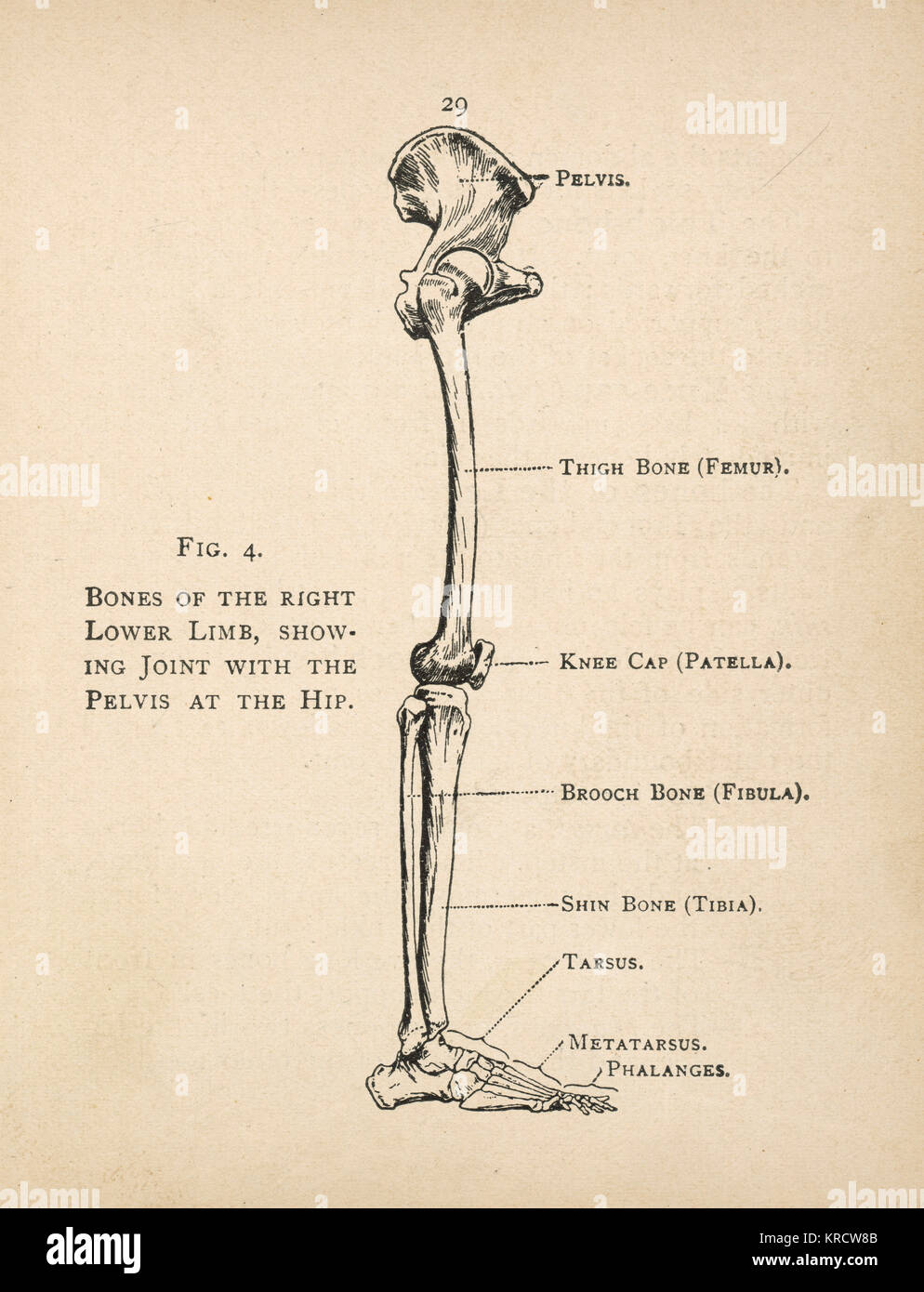
...BrendaLou's Blog...: Phalange...that's what I'm talking about! from 2.bp.blogspot.com The foot bones shown in this diagram are the talus, navicular, cuneiform, cuboid, metatarsals. Your leg bones are very large and strong to help support the weight of your body. Quizzes on human skeletal system anatomy, bone anatomy, and bone markings. The bones involved in it, however, are only the femur and the tibia, although the smaller bone of the leg, the fibula, is carried along in the movements of flexion, extension, and slight rotation that this joint. At the microscopic level, this hard outer. Learn how to draw the femur, patella, tibia, and fibula in this lesson! Bones of the leg and foot, lower leg bone anatomy, leg bones anatomy, leg muscles, leg bones diagram, leg bone structure, leg anatomy muscles, parts of the lower leg. The human leg consists of 8 bones, 4 per leg.
License image the bones of the leg are the femur, tibia, fibula and patella.
Use the leg bones diagrams to learn the names of the leg bones. Learn how to draw the femur, patella, tibia, and fibula in this lesson! Most bones (particularly the long bones of the arms and legs — which make up the appendicular skeleton) have a hard outer shell known as cortical bone. At the microscopic level, this hard outer. The human leg consists of 8 bones, 4 per leg. Health diagram bone skeleton leg knee science anchor chart human human body. Diagram and names of leg bones, diagram of foot and leg bones, diagram of leg bones, diagram of lower leg related posts of diagram of leg bones. A leg bone is a bone found in the leg. They allow you to move and provide support for your upper body. This bright worksheet helps your child bring these technical terms down to size. Skeleton leg ankle joints and toe phalanges, cuboid, metatarsal, navicular and cuneiform bones, hand drawn dorsal view of foot. The bone that goes from your pelvis to your knee is called the femur (say: Quizzes on human skeletal system anatomy, bone anatomy, and bone markings.
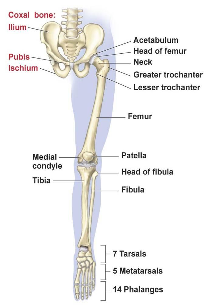
Learn vocabulary, terms and more with flashcards, games and other study tools. The foot bones shown in this diagram are the talus, navicular, cuneiform, cuboid, metatarsals and calcaneus. Learn how to draw the femur, patella, tibia, and fibula in this lesson! Use the leg bones diagrams to learn the names of the leg bones. The bones of the leg are the femur, tibia, fibula and patella.
Human Foot Anatomy Stock Photos & Human Foot Anatomy Stock Images - Alamy from c8.alamy.com Human foot bones anatomy sketch of orthopedics medicine. Synovial joints are often supported and reinforced by surrounding ligaments, which limit movement to prevent injury. The human leg consists of 8 bones, 4 per leg. Diagram and names of leg bones, diagram of foot and leg bones, diagram of leg bones, diagram of lower leg related posts of diagram of leg bones. Includes leg (femur, tibia, patella, and fibula) and foot (tarsals and digits) bones. The foot bones shown in this diagram are the talus, navicular, cuneiform, cuboid, metatarsals. These can include any the following: However, the definition in human anatomy refers only to the section of the lower limb extending from the knee to.
At the microscopic level, this hard outer.
Human foot bones anatomy sketch of orthopedics medicine. Learn the bones of the body with skeletal system quizzes. You'll learn about the muscles, bones, and other structures of each area of the leg. Your leg bones are very large and strong to help support the weight of your body. At the microscopic level, this hard outer. Start learning with our skeleton diagrams, bone labeling exercises and skeletal system quizzes! A leg bone is a bone found in the leg. Bones in the human bodies and names. Health diagram bone skeleton leg knee science anchor chart human human body. Includes leg (femur, tibia, patella, and fibula) and foot (tarsals and digits) bones. Skeleton leg ankle joints and toe phalanges, cuboid, metatarsal, navicular and cuneiform bones, hand drawn dorsal view of foot. High resolution textures and displacement included. The bones and joints in the feet experience wear and tear, so conditions that cause damage to the it is usually the result of a muscle imbalance when the long muscles of the lower leg overpower the.
Learn vocabulary, terms and more with flashcards, games and other study tools. Visit kenhub for more skeletal system quizzes. However, the definition in human anatomy refers only to the section of the lower limb extending from the knee to. Your legs are two of your most important body parts. You'll learn about the muscles, bones, and other structures of each area of the leg.
Lower Limb: Bones, Muscles, Joints & Nerves » How To Relief from www.howtorelief.com The bone that goes from your pelvis to your knee is called the femur (say: Quizzes on human skeletal system anatomy, bone anatomy, and bone markings. At the same time, the bones and joints of the leg and foot must be strong enough to support the body's weight while remaining flexible enough for movement and balance. Bone surfaces at synovial joints are protected by a coating of articular cartilage. Health diagram bone skeleton leg knee science anchor chart human human body. High resolution textures and displacement included. These simple labelled diagrams of the bones of the lower legs and feet and the bones of the arms and hands this diagram shows the skeletal structure of the leg (anterior view) and foot (dorsal view). Interactive anatomical atlas of the head, brain, and neck based on anatomical diagrams and ct and mri medical imaging exams.
Your leg bones are very large and strong to help support the weight of your body.
The foot bones shown in this diagram are the talus, navicular, cuneiform, cuboid, metatarsals. Start learning with our skeleton diagrams, bone labeling exercises and skeletal system quizzes! Interactive anatomical atlas of the head, brain, and neck based on anatomical diagrams and ct and mri medical imaging exams. Your legs are two of your most important body parts. Time to jump right into the biggest and strongest bones in the human body. The foot bones shown in this diagram are the talus, navicular, cuneiform, cuboid, metatarsals and calcaneus. At the same time, the bones and joints of the leg and foot must be strong enough to support the body's weight while remaining flexible enough for movement and balance. However, the definition in human anatomy refers only to the section of the lower limb extending from the knee to. A leg bone is a bone found in the leg. Bones in the human bodies and names. The foot bones shown in this diagram are the talus, navicular, cuneiform, cuboid. The bone that goes from your pelvis to your knee is called the femur (say: Your leg bones are very large and strong to help support the weight of your body.
Share :
Post a Comment
for "Leg Bones Diagram : Forever Horses: Anatomy of the Equine Hindleg"


Post a Comment for "Leg Bones Diagram : Forever Horses: Anatomy of the Equine Hindleg"